
Home | Search | About OC | OC Masterclass Training | Course Schedule | Registration | About Dr. Chan | Doctor Education | Patient Education | Finding a GNM Dentist | Scientific Truth | Dr. Chan’s Articles | Dr. Chan’s Blog Notes | GNM Dentistry | Contact Us
![]()


by James F. Garry, D.D.S.
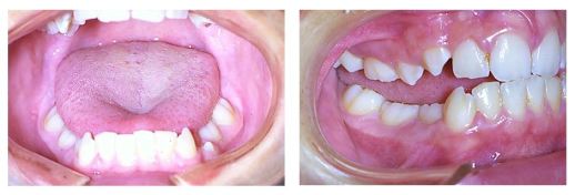
Harvold, in his research with primates, demonstrated that a reduction of tongue size is followed by a corresponding collapse of the dental arches. He found that the tongue could change shape with a plastic insert in the palate. This demonstrated that the shape of the tongue partially depends on its contact with surrounding structures. He found that when a tongue is carried into a new position the distribution of its forces will be altered against the teeth for several months. The teeth, in turn, changed position in response to the new force system with concomitant dental arch changes. Harvold concluded that the extent to which the tongue alters its shape to fit the dental arches depends upon the relative significance of the particular sensation of tooth contact compared with the other sensory inputs. The proprioceptive response of the dorsum of the tongue from hypertrophied adenoids and/or enlarged tonsils may elicit dominant sensory stimuli resulting in a non-physiologic position of the tongue within the dental arches. It has been the author’s clinical experience that all chronic mouthbreathers develop a malocclusion.
Reference: Harvold BP: The Activator inINterceptive Orthopedics, St. Louis: C.V. Mosby Co.;1964;57-63.
Waldeyers Ring
Despite protestations from pediatricians, clinical investigators have failed to demonstrate significant changes in the immunologic status of patients following tonsillectomy and adenoidectomy.


Palatine Tonsils
Almond shaped masses located between the anterior pillars (palatoglossal muscles) and posterior pillars (palatopharyngeal muscles).


Lingual Tonsils
Collection of lymph follicles located at the root of the tongue.


Adenoids
The soft palate has been displaced superiorly with a retractor displaying a large mass of adenoid tissue on the posterior pharyngeal wall of the nasopharynx.


Adult Nasopharynx
View of the adult nasopharynx taken from one of the nasal passages.


Lateral X-ray of Nasopharynx Showing Adenoid Hypertrophy


Acute Hemorrhagic Tonsillitis
Increased bacterial activity produces intense superficial necrosis and ulceration of the mucosa.


Hypertrophy of Palatine Tonsils
Shows marked erythema and edema of the retrotonsillar tissue including the soft palate.
Note the wide deep crypts.


Special Note: Distributing and use of any of these photos to other websites or presentations is considered stealing intellectual property. No permission is granted to anyone accept Dr. Clayton A. Chan who was granted permission directly by Dr. James F. Garry’s son Ron. The originator and author of these images is shared on this site in honor of Dr. Jim to preserve his legacy and a means to encourage other TMJ pain sufferers and dentists hope and better understanding from this site. Copyright permission was given only to Dr. Clayton A. Chan for use on the Occlusion Connections website and no others. Please respect the rights and privacy of Dr. James F. Garry and family members.
Copyright © 2016 Occlusion Connections™ All rights reserved.
___________________________
Read more on:
- Airway Obstruction: Environmental Influences
- Dr. James F. Garry – Biography Dr. James F. Garry – Biography
- The Legacy of James F. Garry, D.D.S. – “The Einstein of Dentistry” – A power point presentation
- “In Loving Memory of My Great Mentor and Dear Friend, Dr. James F. Garry.”
- Upper Airway Obstruction and Upper Airway Deformaties – Part 1
- Upper Airway Obstruction and Upper Airway Deformaties – Part 2
- Defining Neuromuscular Dentistry
- Defining Gneuromuscular Dentistry
- Gneuromuscular vs.Neuromuscular Dentistry
- Computerized Electro-Diagnostic Instrumentation
- Who Are the GNM Dentists?
![]()

9061 West Post Road, Las Vegas, Nevada 89148 United States Telephone: (702) 271-2950
![]()

Leader in Gneuromuscular and Neuromuscular Dentistry